
Why some heart attacks happen in arteries that never looked “blocked”

Why some heart attacks happen in arteries that never looked “blocked”
A normal stress test or “clean” angiogram can reduce concern for major obstructive disease, but it does not completely eliminate the risk of a future heart attack. Here’s the type of plaque behind many of them — and why standard testing can miss it.
A 55-year-old with a clean stress test six months ago walks into the ER with chest pain. By the standard definition, his arteries weren’t “blocked” — but he’s having a heart attack anyway. It’s one of the most common scenarios in cardiology, and the explanation has more to do with what’s inside the artery wall than how wide the opening is.
Cardiac CT with AI plaque analysis in Midtown — results in 48 hours.
What “blocked” actually means
Most traditional cardiac tests — stress tests, EKGs, some catheter-based angiograms — assess whether an artery is narrowed enough to limit blood flow. The clinical threshold is usually around 70 percent narrowing; below that, the test typically reads as “non-significant.” That cutoff makes sense for one question (is the artery so blocked it’s starving the heart during exercise?) but it doesn’t answer a different, equally important one: what is the plaque actually made of, and is it likely to behave dangerously?
What actually ruptures
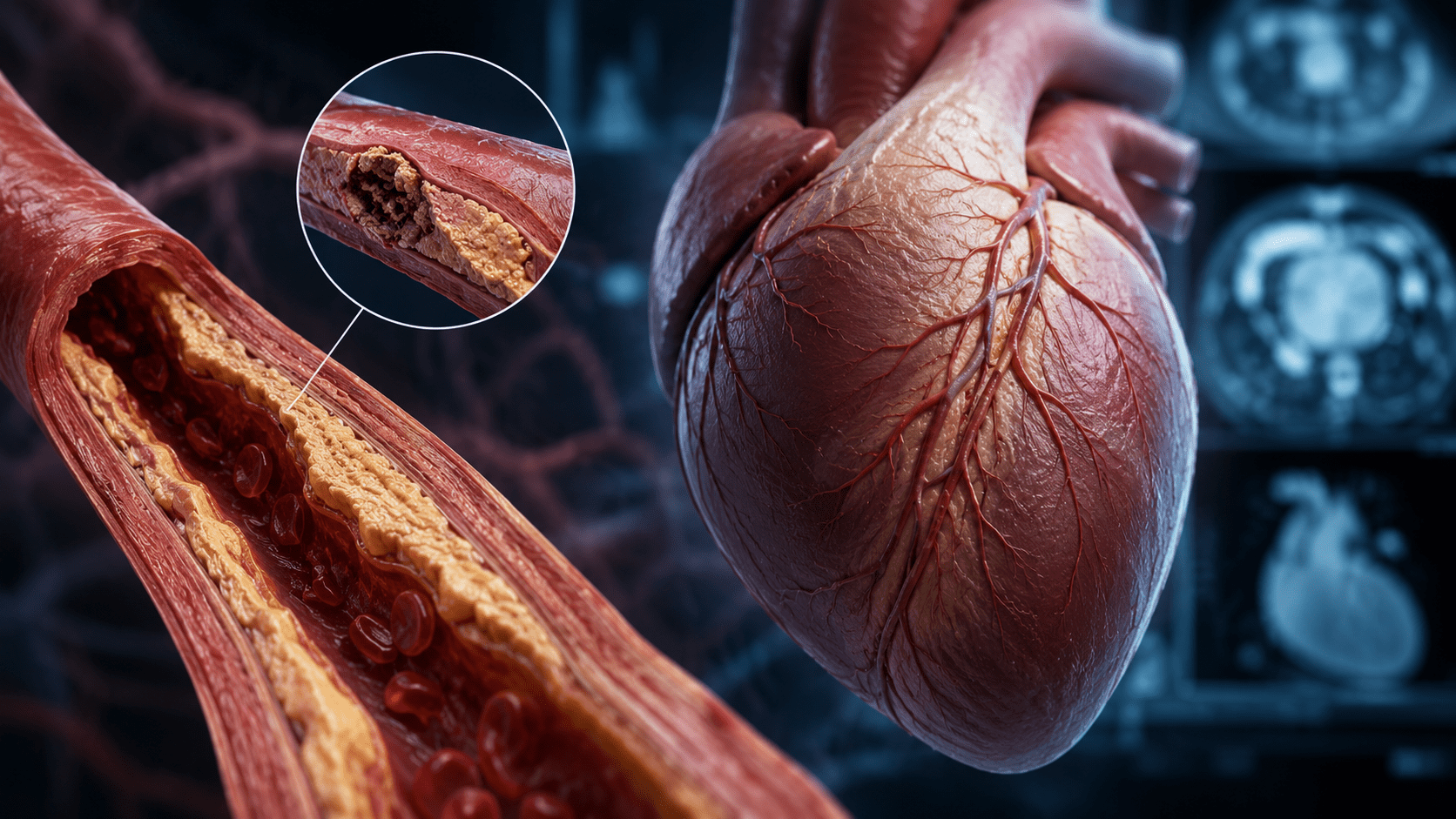
Research has shown that many heart attacks can originate from plaques that were not previously causing the most severe narrowing. They originate from plaques that can appear mild on traditional imaging but have features commonly associated with instability — soft, non-calcified composition, a thin fibrous cap, inflammation, positive remodeling. When one of these plaques ruptures, the body forms a clot at the site, and that clot is what closes the artery acutely. The original narrowing might have been thirty or forty percent. In these cases, plaque composition and stability may play a major role alongside the degree of narrowing.
The Bottom Line
Heart attack risk isn’t only about how narrow an artery is. It’s also about what the plaque is made of. Standard tests measure flow; advanced imaging adds composition to the picture.
What to do about it
For patients with cardiovascular concerns or a strong family history, a coronary CT angiogram is one of the most direct noninvasive ways to evaluate both vessel narrowing and plaque composition — not just whether plaque is there, but what kind. Adding Cleerly AI plaque analysis provides a detailed assessment of plaque burden and plaque features that have been associated with higher cardiovascular risk in clinical studies. At CPAI, both are flat-fee, scanned on a 640-slice open-ring system, and reported within 48 hours by board-certified cardiac radiologists. No referral is required for most patients.
See what’s inside the plaque, not just whether it’s there.
Book a cardiac CT in Midtown Manhattan. Most appointments within the week, reports in 48 hours — evenings, weekends, and Sundays available.
Or call (212) 363-7315