Brain PET/CT: When MRI Shows Structure But Metabolic Mapping Reveals the Answer
Brain PET/CT: When MRI Shows Structure But Metabolic Mapping Reveals the Answer
Your MRI comes back “unremarkable.” The radiologist sees no tumors, no obvious lesions, no structural abnormalities. Yet symptoms persist—memory lapses, confusion, personality changes, unexplained neurological decline.
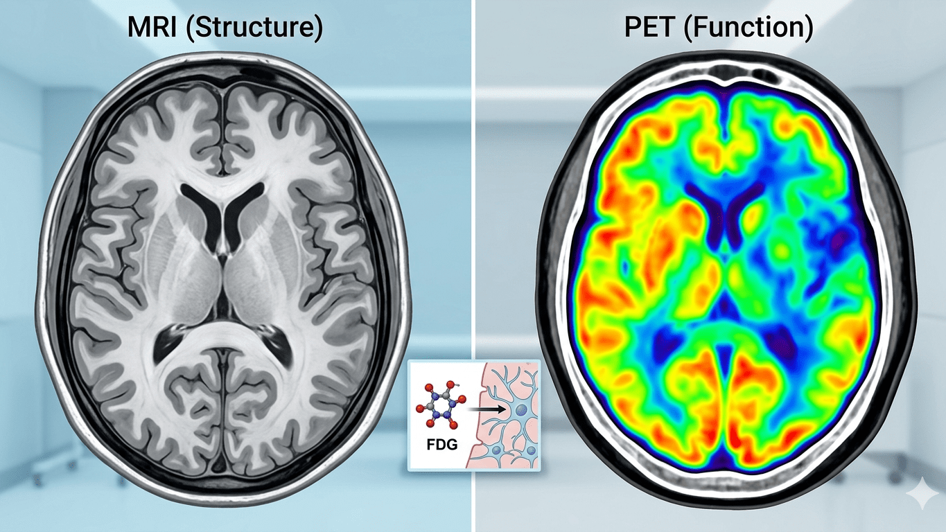
The problem? MRI shows brain anatomy. It doesn’t show brain function.
Brain PET/CT imaging fills this critical gap. While MRI visualizes structure, PET reveals how brain cells are functioning at the metabolic level—often detecting disease years before structural changes become visible.
Metabolic Changes Precede Structural Changes
Diseased or degenerating brain cells consume less glucose than healthy tissue. PET imaging detects these metabolic changes long before tissue loss, atrophy, or structural abnormalities appear on MRI—enabling earlier diagnosis when intervention matters most.
What Brain PET Actually Measures
Brain PET imaging uses a radiotracer—typically FDG (fluorodeoxyglucose), a glucose analog—to map how actively different brain regions are metabolizing energy.
Healthy brain tissue uses glucose steadily and uniformly. Diseased, degenerating, or damaged tissue shows reduced metabolic activity—appearing as “dim” regions on PET scans while still looking normal on MRI.
This metabolic signature is what makes PET invaluable for conditions where structural imaging provides insufficient answers.
When PET Answers Questions MRI Cannot
Dementia Differential Diagnosis
Many forms of dementia—Alzheimer’s disease, Lewy body dementia, frontotemporal dementia, vascular dementia—appear similar clinically and often show minimal early changes on MRI. But each produces distinct metabolic patterns on PET.
Alzheimer’s shows characteristic hypometabolism in posterior parietal and temporal regions. Lewy body dementia affects occipital lobes. Frontotemporal dementia primarily reduces metabolism in frontal and anterior temporal areas. PET identifies these patterns when MRI remains inconclusive.
Brain Tumor Monitoring
After surgery or radiation therapy, MRI often cannot distinguish between residual active tumor and post-treatment scar tissue—both appear as masses on structural imaging. PET resolves this ambiguity by showing metabolic activity. Active tumor metabolizes glucose aggressively. Scar tissue does not.
Refractory Seizure Localization
For patients with medication-resistant epilepsy, identifying the seizure focus is critical for surgical planning. PET reveals hypometabolic regions between seizures that correspond to seizure origins—guidance that structural imaging cannot provide.
Unexplained Cognitive Decline
When memory loss, confusion, or cognitive changes occur without clear explanation and MRI shows no abnormalities, PET provides metabolic insight that can identify neurodegenerative processes in their earliest stages.
The Technology: Digital PET at 2.9mm Resolution
At Central Park Advanced Imaging, we use the United Imaging uMI 550 digital PET/CT system—one of the highest-resolution brain imaging platforms available.
Digital PET delivers 2.9mm resolution with AI motion correction, detecting subtle metabolic changes that older analog systems miss. The entire brain scan completes in as little as 8 minutes, minimizing patient discomfort and motion artifacts.
This technology enables quantitative analysis—standardized uptake value (SUV) measurements that allow objective comparison and longitudinal tracking over time.
The Brain PET Experience
The process is straightforward. After fasting for 4-6 hours (water permitted), you receive a small injection of FDG tracer. You rest quietly for 30-60 minutes while the tracer circulates and is absorbed by brain tissue.
The scan itself takes 8-15 minutes. You lie comfortably on the scanning table—no enclosed space, no loud noises like MRI. The entire appointment, including preparation, takes approximately 2-2.5 hours.
Results are interpreted by board-certified radiologists specializing in nuclear medicine and delivered to your physician within 24-48 hours.
For complete program details, visit our Brain PET/CT program page.
Call to Action
When structural imaging leaves questions unanswered, brain PET/CT provides metabolic insight that changes diagnosis and treatment. Schedule your consultation or call (212) 363-7315 to learn more. Contact us with questions about brain PET imaging.