This Is Not a Standard CT Scan: Inside CPAI’s United Imaging 640-Slice Technology
This Is Not a Standard CT Scan: Inside CPAI’s United Imaging 640-Slice Technology
When your doctor orders a CT scan, you probably assume all CT scanners are roughly the same. You’d be wrong. The difference between a standard 64-slice CT and Central Park Advanced Imaging’s United Imaging uCT Atlas 640-slice system isn’t incremental—it’s transformative. It’s the difference between a snapshot and a complete story, between guesswork and certainty, between motion blur and crystal clarity.
Most imaging centers in New York still operate 64- or 128-slice CT scanners—technology that’s been the standard for years. At CPAI, we’ve moved beyond standard. Our 640-slice system captures entire organs in a single 0.25-second rotation, eliminating the motion artifacts that compromise image quality and diagnostic confidence. This isn’t just a better CT scan. It’s a fundamentally different approach to seeing inside the human body.
Why Slice Count Matters
The number of slices in a CT scanner determines how much of your body can be imaged simultaneously. A 64-slice scanner captures 64 cross-sectional images per rotation. Our 640-slice system captures ten times that amount—providing 160mm of z-axis coverage in one rotation. That means we can image your entire heart, or your entire brain, in the time it takes your heart to beat once.
The Problem With Standard CT Scans
Traditional CT scanners face a fundamental challenge: organs move. Your heart beats. Your lungs expand. Even your abdominal organs shift with respiration. Standard 64- or 128-slice scanners must take multiple rotations to capture a complete image of moving organs, which means they’re stitching together images taken at different moments in time. The result? Motion artifacts, blurred edges, and diagnostic uncertainty.
For cardiac imaging, this limitation is particularly problematic. A beating heart doesn’t hold still, and traditional scanners struggle to freeze that motion. Radiologists often see blurred coronary arteries, making it difficult to assess blockages or plaque buildup with confidence. Patients are sometimes asked to return for repeat scans, or doctors must make clinical decisions based on suboptimal images.
How 640-Slice Technology Changes Everything
CPAI’s uCT Atlas system solves this problem through sheer computational power and engineering precision. With 640 detector rows and 0.25-second rotation speed, the scanner captures 160mm of anatomical coverage in a fraction of a heartbeat. This single-rotation acquisition means motion-free imaging of the heart, brain, lungs, and other organs—no stitching, no artifacts, no compromises.
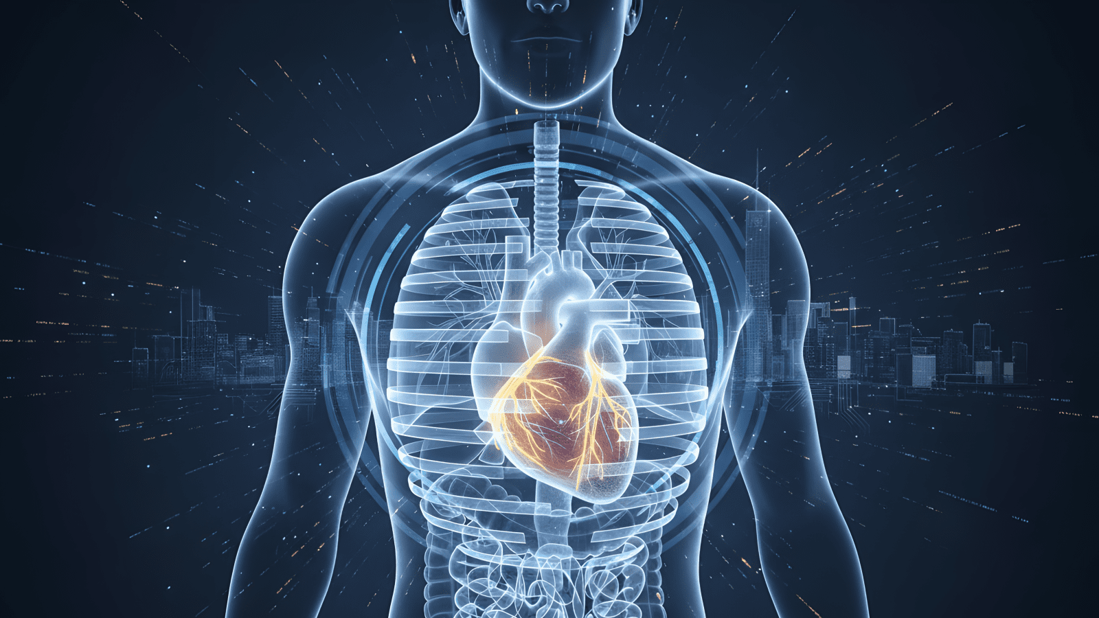
Single-Heartbeat Cardiac Imaging
Cardiac CT angiography requires visualizing the coronary arteries with exceptional clarity. The uCT Atlas captures your entire heart—all four chambers, all major vessels, all coronary branches—in one rotation, frozen in time during a single heartbeat. This produces images sharp enough to identify even subtle plaque deposits or early-stage blockages that standard scanners might miss or mischaracterize.
For patients with irregular heartbeats or difficulty holding their breath, this capability is transformative. The speed and coverage eliminate the variables that degrade image quality on traditional systems, providing diagnostic clarity regardless of heart rate or respiratory cooperation.
Trauma Imaging When Seconds Count
In emergency situations, speed isn’t a luxury—it’s a necessity. The 640-slice system’s ability to image the entire chest, abdomen, and pelvis in seconds makes it invaluable for trauma evaluation. Radiologists can assess multiple organ systems simultaneously, identifying internal bleeding, fractures, or organ damage without delay. This comprehensive coverage in minimal time supports faster diagnosis and more confident emergency decision-making.
AI-Powered Precision With Lower Radiation
Advanced hardware is only part of the story. The uCT Atlas integrates Deep IR reconstruction—a dual-layer AI system that combines physics-based modeling with neural networks to reduce noise and enhance image quality. This means diagnostic clarity at lower radiation doses, an especially important consideration for patients requiring repeated imaging over time.
The scanner’s uAI Vision workflow suite includes automated patient positioning, intelligent scan range selection, and organ-based dose optimization. These AI-driven features ensure every scan is tailored to your anatomy and clinical needs, maximizing diagnostic value while minimizing radiation exposure.
Dual-Energy Imaging: Seeing Beyond Anatomy
The uCT Atlas offers dual-energy CT, which uses two different X-ray energy levels to differentiate tissue types based on their material composition. This allows radiologists to distinguish between iodine contrast and calcium deposits, create virtual non-contrast images, and even identify kidney stone composition—all from a single scan.
Designed for Real Patients, Not Just Ideal Cases
Advanced technology means nothing if patients can’t fit comfortably or safely inside the scanner. The uCT Atlas features an 82cm ultra-wide bore—significantly larger than the 70cm standard—and a table capacity of 318 kg (approximately 700 pounds). This isn’t about accommodating edge cases; it’s about recognizing that real patients come in all sizes, and everyone deserves access to the most advanced imaging available.
The wider bore also reduces claustrophobia and anxiety, making the scan experience more comfortable for patients who might otherwise struggle in tight spaces. Faster scan times mean less time in the machine, further improving the patient experience without sacrificing image quality.
What This Means for Your Health
Technology specifications matter, but what matters more is what they enable: earlier detection, greater diagnostic confidence, and more personalized treatment planning. The 640-slice system’s ability to capture motion-free images of moving organs means radiologists can identify findings that might be missed or obscured on standard scanners. For cardiac patients, this could mean detecting coronary artery disease before it causes symptoms. For lung screening patients, it means clearer visualization of small nodules. For anyone undergoing imaging, it means results you and your doctor can trust.
At CPAI, the uCT Atlas isn’t used in isolation. Our board-certified radiologists review every study on-site, often delivering results within 24 hours. Same-week appointments mean you don’t wait weeks for access to advanced imaging. This combination—cutting-edge technology, expert interpretation, and concierge-level service—creates an experience that prioritizes your time, comfort, and diagnostic certainty.
Beyond Standard Care
Many imaging centers treat CT scans as a commodity service, prioritizing volume over precision. At CPAI, we’ve made a different choice. By investing in the most advanced CT technology available—systems typically found only at major academic medical centers—we’re able to offer Manhattan patients imaging that goes beyond standard care.
Whether you need coronary artery evaluation, lung screening, abdominal imaging, or comprehensive trauma assessment, the difference between adequate imaging and exceptional imaging can be significant. Motion-free cardiac imaging reveals details that blurred images cannot. Low-dose protocols protect your long-term health without compromising diagnostic quality. Single-rotation coverage means faster scans and less time away from your life.
The Choice Is Yours
Not all CT scans are created equal. The 640-slice difference isn’t marketing language—it’s a fundamental shift in what’s possible with computed tomography. At Central Park Advanced Imaging, this technology is available now, with same-week appointments and results that arrive when you need them. You deserve imaging that matches the complexity of your health, interpreted by radiologists who prioritize accuracy over speed, delivered in an environment designed around your comfort and convenience.
Take control of your diagnostic journey with imaging technology that doesn’t compromise. Schedule your CT scan at CPAI today and experience the clarity that comes from truly advanced imaging. Have questions about whether our 640-slice CT is right for your needs? Contact our team—we’re here to help you make informed decisions about your health.