The Difference Between 1.5T and 3T MRI: Seeing the Unseen
The Difference Between 1.5T and 3T MRI: Seeing the Unseen
When scheduling an MRI, you might encounter the terms “1.5T” and “3T”—references to magnetic field strength measured in Tesla units. This technical detail matters more than most patients realize. The difference between these field strengths fundamentally affects what can be detected, how quickly scans complete, and which subtle abnormalities become visible.
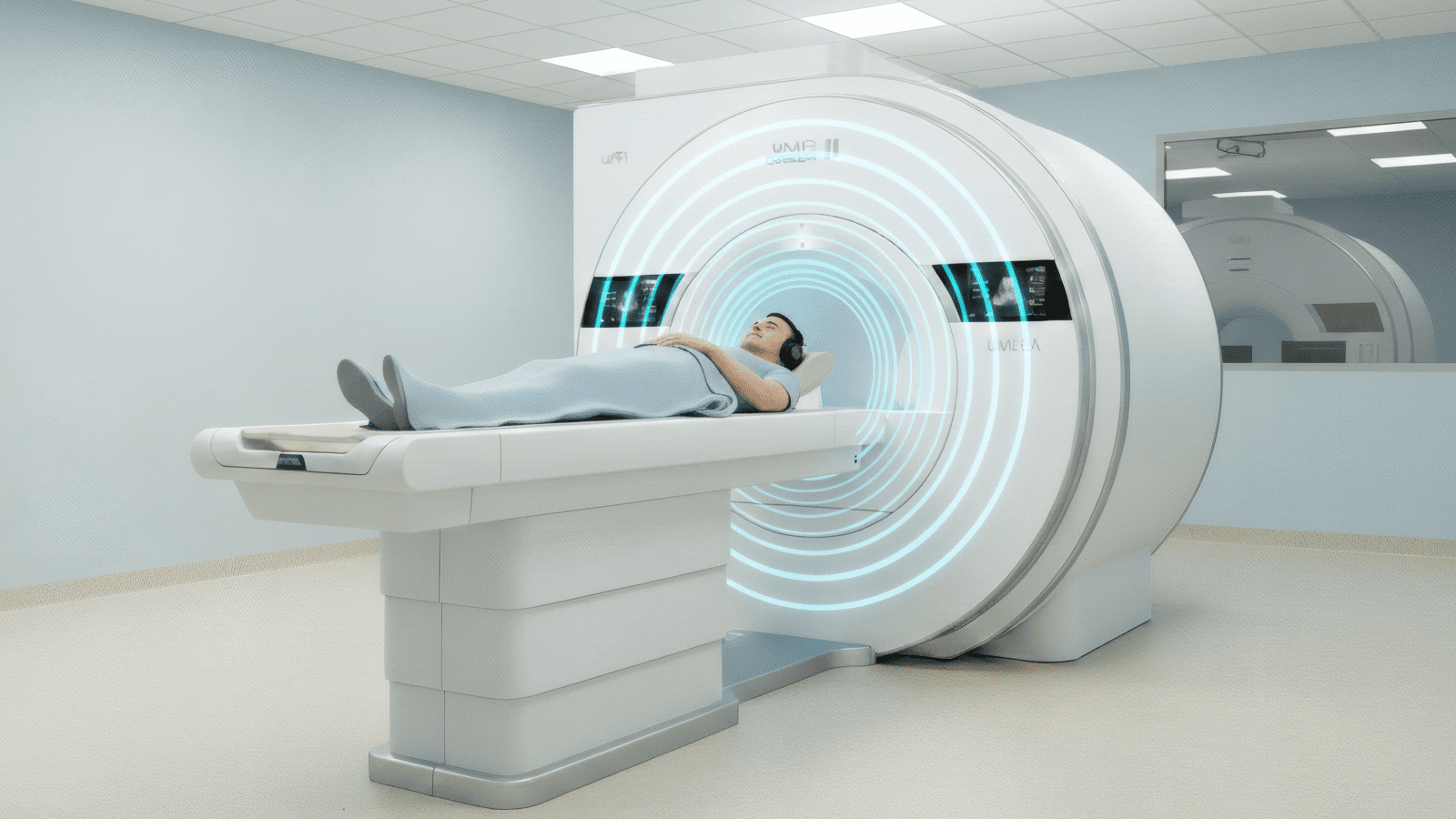
At Central Park Advanced Imaging, our uMR Omega 3T system provides double the magnetic field strength of standard 1.5T scanners. Understanding what this means in practical terms helps you appreciate the technology’s capabilities and why we’ve invested in Manhattan’s most advanced MRI.
Understanding Tesla
Tesla measures magnetic field strength. Earth’s magnetic field is approximately 0.00005 Tesla. A 1.5T MRI magnet is 30,000 times stronger than Earth’s field. A 3T system doubles that strength to 60,000 times Earth’s field—generating the powerful signals needed for detailed imaging.
The Physics: Why Stronger Magnets Matter
MRI works by aligning hydrogen atoms in your body with a strong magnetic field, then using radiofrequency pulses to disturb that alignment. As atoms return to their aligned state, they emit signals that scanners detect and convert into images. Stronger magnetic fields align more hydrogen atoms, generating stronger signals that translate to better image quality.
Signal-to-Noise Ratio: The Core Advantage
- Theoretical Improvement: Signal increases proportional to the square of field strength—theoretically doubling signal-to-noise ratio (SNR) from 1.5T to 3T.
- Practical Reality: Tissue properties reduce actual SNR improvement to 30-60%, still delivering substantially better spatial resolution and faster scanning.
- Trading SNR: The improved signal can be “spent” on higher resolution images, faster acquisition, or thinner slices—flexibility that transforms clinical capabilities.
Image Quality: Seeing Finer Detail
The SNR advantage of 3T translates directly into superior image quality. Radiologists consistently rate 3T images higher for visualization of anatomical structures and pathological findings across nearly all body regions.
What 3T Reveals
- Spatial Resolution: 3T enables slice thickness below 1mm versus 2-3mm typically on 1.5T—revealing structures too small to resolve at lower field strength.
- Cartilage Visualization: Studies demonstrate higher diagnostic confidence at 3T, with better sensitivity for detecting subtle cartilage lesions before they progress.
- Soft Tissue Contrast: Enhanced signal differentiation between tissue types improves detection of small tumors, inflammatory changes, or early degeneration.
- Edge Definition: Sharper delineation of boundaries between structures—critical for surgical planning and precise diagnosis.
“The difference between 1.5T and 3T isn’t just technical—it’s about detecting disease at earlier, more treatable stages when intervention can make the greatest impact.”
Speed Advantages: Shorter Scan Times
The SNR improvement at 3T enables significantly faster image acquisition without compromising diagnostic quality. Studies show 30-50% reduction in scan time for equivalent image quality compared to 1.5T protocols.
Time Savings That Matter
- Brain Imaging: Complete neuroimaging exams achievable in under 10 minutes at 3T versus 20-30 minutes at 1.5T.
- Joint Studies: Full knee examination completed in 5-8 minutes rather than 15-20 minutes.
- Patient Comfort: Faster scans particularly benefit claustrophobic patients, children, individuals with pain, and emergency situations requiring rapid diagnosis.
Clinical Applications: Where 3T Excels
While 3T offers advantages across most imaging applications, certain clinical scenarios particularly benefit from the higher field strength.
Neuroimaging Excellence
- Multiple Sclerosis: Superior detection of small demyelinating lesions—consensus guidelines now recommend 3T for MS diagnosis and monitoring.
- Tumor Characterization: Better delineation of tumor margins, improved detection of small metastases.
- Functional Imaging: Enhanced signal makes functional MRI more sensitive for mapping brain activity.
Prostate Imaging Precision
- Lesion Detection: PI-RADS guidelines prefer 3T for prostate MRI, citing superior resolution and contrast.
- No Endorectal Coil: Higher inherent signal at 3T often eliminates need for uncomfortable endorectal coils still required at 1.5T.
- Multiparametric Protocols: Enhanced dynamic contrast imaging and diffusion-weighted sequences guide targeted biopsies.
Musculoskeletal Detail
- Cartilage Assessment: Detection of early degenerative changes before they become symptomatic.
- Ligament Tears: Better visualization of partial tears and improved characterization of injury severity.
- Sports Medicine: Detailed assessment of complex injuries involving multiple tissue types.
The 3T Advantage at CPAI
Our uMR Omega 3T combines higher field strength with innovations addressing traditional 3T challenges—delivering superior image quality with enhanced patient comfort.
Why Our 3T Is Different
- 75cm Ultra-Wide Bore: Widest 3T MRI available worldwide—superior comfort without sacrificing field strength advantages.
- QuietScan Technology: Up to 80% noise reduction—addressing a primary 3T disadvantage.
- uAI DeepRecon: Deep learning reconstruction enables 80% faster scanning with 44% SNR improvement—multiplying 3T’s inherent benefits.
- 310 kg Table Capacity: Accommodates all body types on a 3T system—accessibility without compromising technology.
When to Choose 3T
Field strength selection depends on clinical indication and diagnostic goals. Understanding when 3T makes the most difference ensures you’re getting the right technology for your situation.
3T Is Particularly Valuable For:
- Complex Anatomy: Brain, spine, joints, and prostate where detail matters most.
- Small Structures: Lesions, tears, or abnormalities requiring high resolution to visualize.
- Follow-Up Imaging: Establishing baseline at 3T provides highest sensitivity for detecting future changes.
- Time Constraints: When rapid diagnosis is critical or patient tolerance is limited.
Experience the Difference
The “unseen” in 3T imaging refers to pathology below 1.5T’s detection threshold—lesions too small, contrast differences too subtle, or structures too detailed for lower field strength visualization. At CPAI, our uMR Omega 3T provides the diagnostic advantages of high-field imaging while addressing comfort, noise, and accessibility concerns.
Whether detecting subtle early disease, characterizing complex pathology, or requiring rapid imaging, our technology delivers superior results. Contact us to learn more about 3T MRI imaging, call us at (212) 363-7315, or book your scan online.