Thyroid & Breast Health: How AI-Powered Ultrasound Improves Accuracy
Thyroid & Breast Health: How AI-Powered Ultrasound Improves Accuracy
Thyroid & Breast Health: How AI-Powered Ultrasound Improves Accuracy
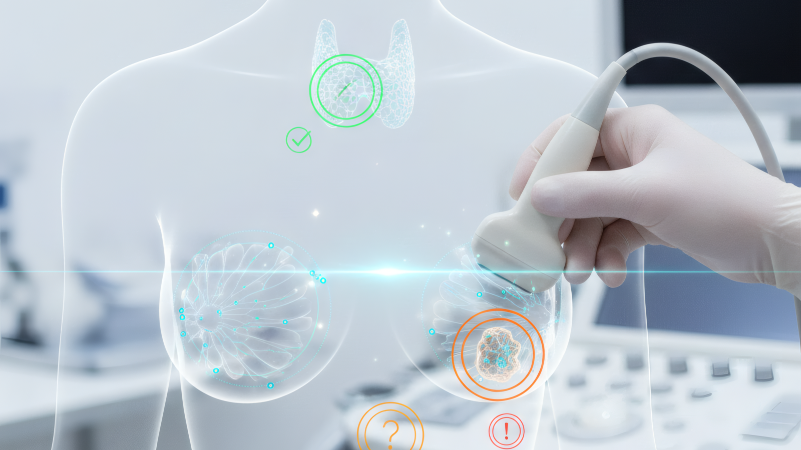
Thyroid nodules and breast findings are common across the population, and most are benign. The clinical challenge has always been distinguishing which findings require closer attention and which can simply be monitored over time.
At Central Park Advanced Imaging, our Samsung RS85 Premium Ultrasound System integrates S-Detect AI technology that standardizes lesion analysis and supports more consistent interpretation. This deep learning algorithm evaluates ultrasound images in real time, characterizing findings according to established clinical frameworks and providing objective insight that complements physician interpretation.
Understanding AI-Assisted Diagnosis
S-Detect does not replace radiologist expertise—it enhances it. The AI evaluates specific image features (such as shape, margins, and internal structure) and applies standardized classification systems for both breast and thyroid imaging. This supports consistency and provides an added layer of analytical confidence.
How AI Improves Diagnostic Accuracy
Traditional ultrasound interpretation can vary based on operator experience and subjective assessment. In borderline cases, this variability may lead to uncertainty or inconsistent recommendations.
S-Detect introduces consistent, structured analysis. Each finding is evaluated using the same criteria every time, supporting reproducible assessments and enhanced interpretive clarity.
What S-Detect Analyzes
- Composition: Differentiates solid, cystic, and mixed findings.
- Echogenicity: Evaluates how a finding reflects ultrasound waves relative to surrounding tissue.
- Shape and Orientation: Assesses structural features used in standardized classification.
- Margins: Reviews border characteristics to support risk stratification.
- Calcifications: Identifies internal echo patterns commonly evaluated in breast and thyroid imaging.
- Vascularity: Assesses blood flow patterns using advanced Doppler techniques.
Clinical Performance and Consistency
Multiple published studies support the clinical reliability of AI-assisted ultrasound across diverse patient populations. These studies demonstrate that AI-supported interpretation enhances consistency and assists radiologists in confidently categorizing findings.
Thyroid Nodule Assessment
- Consistent Classification: AI standardizes TI-RADS assessment for improved reproducibility.
- Reliable Benign Identification: AI support helps reduce unnecessary follow-up in clearly benign patterns.
- Improved Reader Confidence: Diagnostic confidence increases when AI and physician interpretation align.
Breast Lesion Evaluation
- BI-RADS Support: S-Detect applies standardized BI-RADS criteria for consistent classification.
- Reader Assistance: AI support enhances specificity and interpretive confidence.
- Efficient Workflow: Improved first-pass assessment supports fewer repeat studies.
Health Tips: When to Consider Ultrasound
Ultrasound offers valuable, real-time insight into thyroid and breast health and is commonly used for both screening and diagnostic evaluation.
Common Thyroid Evaluation Scenarios
- Personal Neck Awareness: Any new or changing neck finding may be evaluated with ultrasound.
- Family Health Awareness: Many patients choose baseline imaging to better understand their thyroid health.
- Previous Imaging Findings: Nodules noted incidentally on CT or MRI are often further evaluated with ultrasound.
- Thyroid Function Changes: Hormonal changes may sometimes be accompanied by structural findings.
- Routine Health Visibility: Some patients choose ultrasound proactively as part of comprehensive wellness screening.
Common Breast Ultrasound Scenarios
- Dense Breast Tissue: Supplemental ultrasound enhances visualization in dense breast patterns.
- Localized Awareness: Any area of concern can be evaluated in real time.
- Personal Health Monitoring: Many women choose ultrasound as part of an ongoing wellness strategy.
- Family Health Awareness: Baseline screening is commonly used in those with family history.
- Mammographic Follow-Up: Ultrasound supports further characterization of mammographic findings.
Preparing for Your AI-Assisted Ultrasound
Preparation is simple, and our team provides clear guidance in advance to ensure a smooth, comfortable visit.
Before Your Thyroid Ultrasound
- Remove Neck Jewelry: Allows optimal probe positioning.
- Wear Open-Neck Clothing: Supports easy access without changing.
- Note Any Symptoms: Share anything you’ve noticed with your technologist.
- Bring Medication Information: Helpful for context.
- Bring Prior Imaging: Prior studies support direct comparison.
Before Your Breast Ultrasound
- Schedule Comfortably: Many patients prefer the week after menstruation when applicable.
- Avoid Lotions: Skin products may interfere with image clarity.
- Note Any Areas of Interest: Share location and timing of anything you’ve noticed.
- Bring Prior Imaging: Mammogram reports and prior studies support better comparison.
- Know Your History: Prior procedures or family history provide helpful context.
Understanding Your Results
S-Detect generates structured image analysis that your radiologist reviews within your complete clinical context. This supports clear, personalized interpretation.
Thyroid Nodule Classification
- Benign Patterns: Typically require routine monitoring or no follow-up.
- Low-Level Suspicion: Often managed with interval observation.
- Moderate Suspicion: May prompt additional evaluation depending on size and clinical context.
- Higher Suspicion: Follow-up recommendations are tailored individually.
Breast Lesion Categories
- Benign Findings: Continue routine wellness screening.
- Probably Benign Patterns: Often followed with short-interval imaging for confirmation.
- Suspicious Findings: Additional evaluation may be recommended for clarity.
- Highly Suspicious Patterns: Prompt follow-up supports comprehensive care planning.
What Happens After Your Scan
Clear communication and continuity are built into every step of your experience.
Results Timeline and Next Steps
- Image Review: Board-certified radiologists review your study promptly.
- Communication: Results are shared clearly through your physician or care team if requested.
- Optional Follow-Up: Some findings invite additional imaging for clarity.
- Monitoring Protocols: Follow-up intervals are personalized to your goals.
- Comparison Studies: AI-assisted analysis supports precise comparison over time.
The Advantage of AI-Assisted Imaging
AI-assisted ultrasound supports objective, reproducible analysis alongside expert physician interpretation. This combination enhances confidence, reduces uncertainty, and supports clearer clinical decision-making.
For patients, this means consistency and clarity. When expert review and AI analysis align, confidence in next steps is strengthened. When they highlight something new, you gain earlier visibility and actionable insight.
Looking Ahead
AI continues evolving as algorithms refine performance through expanding clinical datasets. As false positives continue decreasing and subtle finding detection continues improving, AI-assisted ultrasound will play an even broader role in preventive imaging.
At CPAI, our Samsung RS85 with S-Detect AI reflects our commitment to bringing the most advanced ultrasound technology into a patient-first, concierge-level environment. Whether you’re pursuing proactive wellness screening or evaluating a specific concern, AI-assisted ultrasound delivers clarity, consistency, and confidence. Schedule your thyroid or breast ultrasound with AI-powered analysis.