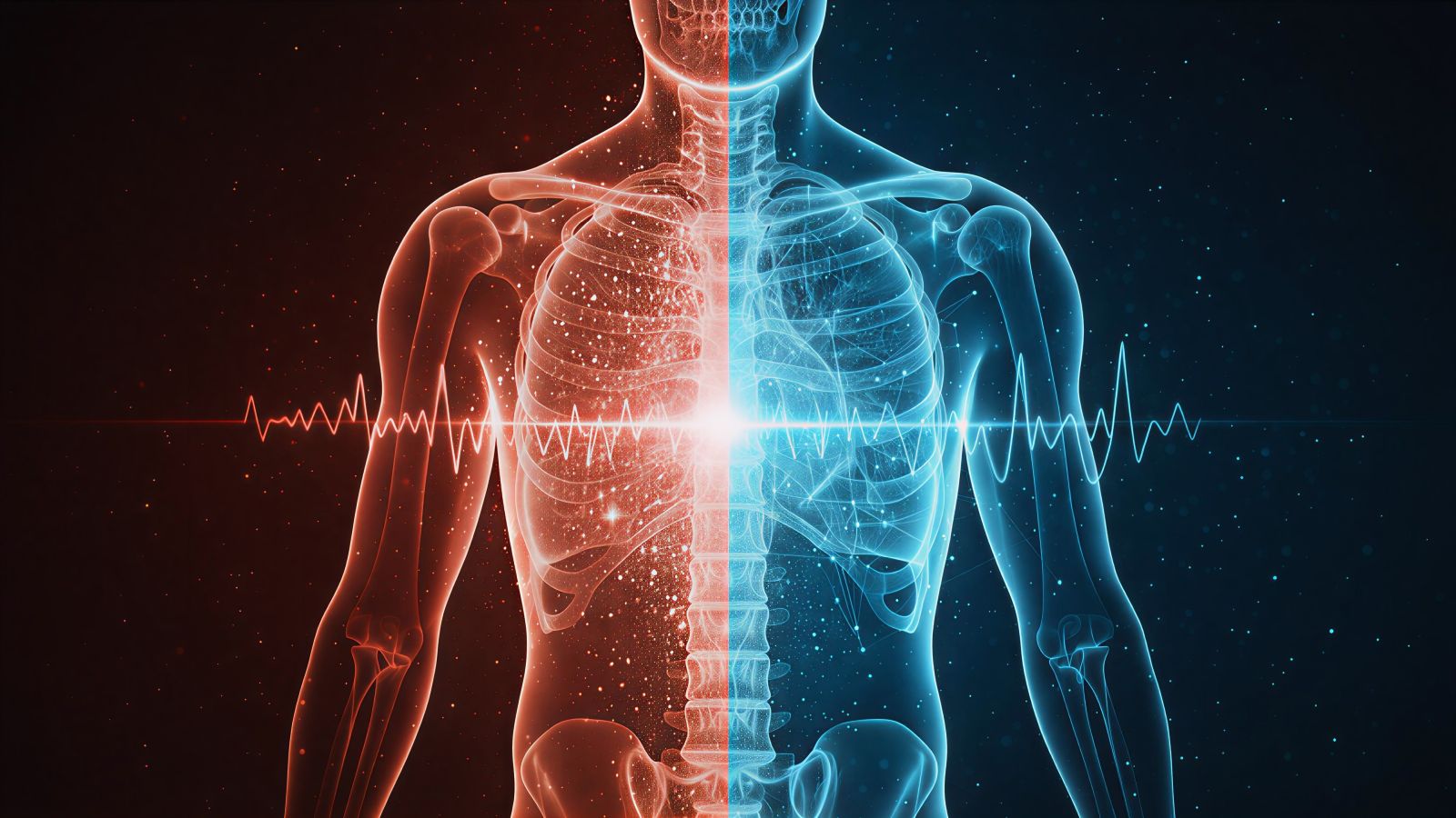
Understanding “Dose Optimization”: How AI Lowers Radiation in CT Scans
Understanding “Dose Optimization”: How AI Lowers Radiation in CT Scans
Understanding “Dose Optimization”: How AI Lowers Radiation in CT Scans
For decades, CT imaging faced a fundamental tradeoff: reducing image noise typically required increasing radiation dose. Clearer images generally meant more X-rays. This relationship shaped CT imaging for years—until artificial intelligence transformed how image quality and dose efficiency are balanced.
At Central Park Advanced Imaging, our United Imaging uCT Atlas 640-Slice CT uses Deep IR reconstruction—an AI-powered technology that supports advanced dose optimization. The result is high-quality diagnostic imaging with meaningfully reduced radiation compared to earlier-generation CT systems, while maintaining the image clarity radiologists rely on.
The Breakthrough That Changes Everything
Deep learning reconstruction uses neural networks trained on large image datasets to distinguish true anatomical signal from random noise. Unlike older methods that relied on smoothing to suppress noise, AI-based reconstruction preserves fine detail while supporting lower-dose imaging across a wide range of protocols.
The Evolution: From Hardware to Intelligence
Traditional CT reconstruction, known as Filtered Back Projection (FBP), was introduced in the 1970s. It remains fast and reliable, but image noise is directly linked to radiation dose—cleaner images require higher exposure.
Iterative reconstruction (IR), introduced in the 2000s, improved upon this by mathematically modeling the imaging process and reducing noise through repeated calculations. While it enhanced dose efficiency, image texture could appear over-smoothed and dose reductions were limited before image quality began to change.
How Deep Learning Reconstruction Works
- Neural Network Training: The system learns from extensive imaging datasets to differentiate true anatomical structures from random noise.
- Real-Time Application: During reconstruction, the trained network processes lower-dose images and intelligently suppresses noise while preserving anatomical detail.
- Natural Image Texture: Deep learning helps maintain familiar image appearance for radiologists while supporting enhanced contrast and edge clarity.
- Dose Optimization: Image quality becomes less dependent on raw X-ray output, allowing greater flexibility in protocol design.
“Deep learning reconstruction adds a powerful new layer to CT imaging by improving how image data is processed and refined, supporting excellent clarity at optimized dose levels.”
The Clinical Evidence: Meaningful Dose Reductions
Multiple published studies demonstrate that deep learning reconstruction supports significant dose optimization compared to traditional reconstruction methods while preserving diagnostic performance. Results vary by exam type, patient size, and protocol, but consistently demonstrate improved noise performance at reduced exposure levels.
Where Dose Reduction Matters Most
- Pediatric Imaging: Dose optimization is especially valuable in pediatric imaging, where minimizing radiation exposure is a priority.
- Serial Monitoring: Patients who undergo repeated CT scans benefit from cumulative dose efficiency over time.
- Lung Screening: Low-dose chest CT protocols are further supported by AI-based reconstruction for enhanced clarity at reduced exposure.
- Younger Adults: Optimization supports long-term dose awareness across a wide range of clinical use cases.
Beyond Reconstruction: Intelligent Workflow Automation
The uCT Atlas integrates AI across the entire scan workflow—not only in reconstruction, but also in automated positioning and protocol selection. The uAI Vision system supports dose optimization at each stage of the scan, helping ensure efficient, consistent image acquisition.
AI-Powered Dose Control Features
- Auto-Positioning: 3D camera guidance supports accurate patient centering for consistent exposure.
- Intelligent Range Selection: AI recognizes anatomical landmarks and helps define optimal scan coverage.
- Organ-Based Modulation: Adaptive exposure adjustment supports sensitive tissue dose management.
- Adaptive Protocols: Technique factors are tailored based on patient size and exam purpose to avoid one-size-fits-all settings.
The Patient Impact: Clarity Without Compromise
Dose optimization does not mean compromised imaging. Studies show that deep learning–reconstructed CT images maintain excellent detectability for a wide range of clinical findings, including subtle pulmonary, abdominal, and musculoskeletal features.
For patients, this means advanced diagnostic clarity with optimized radiation exposure. Each scan is carefully tailored to deliver the information needed while supporting long-term dose awareness.
Looking Ahead
The integration of AI into CT imaging represents a meaningful evolution in how image quality and radiation efficiency work together. As neural networks continue to improve through ongoing clinical data refinement, dose optimization strategies will continue advancing as well.
At CPAI, we’re committed to providing imaging technology that balances diagnostic confidence with optimized radiation management. Our AI-powered CT systems support both exceptional image clarity and thoughtful dose efficiency. Contact us to learn more about how our advanced CT technology supports confident, informed imaging.